
The Truth About Oxalates in the Body: From Diet to Disease
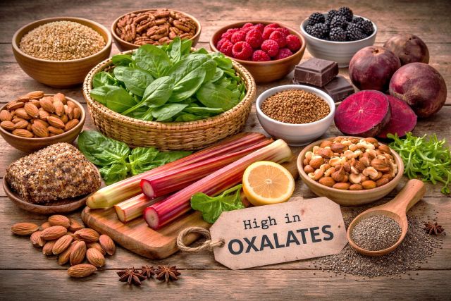
Oxalates serve a protective function in nature. Plants use them as a natural defense against pests by forming sharp-edged crystals that can damage the tissues of insects.
Oxalates are also naturally present in the human body. Some are produced as part of normal metabolism, while others come from the foods we eat. However, their effects depend not only on how many oxalates are consumed. An important factor is how much of them are absorbed in the intestines and how effectively the body is able to eliminate them.
Some people are at a higher risk of oxalate accumulation. This may be due to inherited factors, intestinal disorders, an imbalance in the gut microbiome, or a high dietary intake of oxalates. In these individuals, even a typical diet may sometimes contribute to health problems.
What Are Oxalates?
Oxalates are salts of oxalic acid that are naturally present in the human body, plants, and many foods.

Calcium oxalate crystals (scanning electron microscopy, approximately 5–20 μm in size).
The key characteristic of oxalates is their ability to bind to certain minerals, particularly calcium and magnesium. This results in the formation of different salts with varying solubility. The most clinically important is calcium oxalate, which is poorly soluble and can form crystals.
As long as oxalates remain dissolved, they can be transported in body fluids and eliminated. However, when they begin to crystallize, they form hard crystals with sharp edges.
It is the formation of these crystals—not the mere presence of oxalates—that can lead to mechanical damage to tissues and trigger inflammation.
It is important to understand that the risk of crystal formation depends on more than just the amount of oxalates present. It is also influenced by mineral levels, the acidity (pH) of the environment, hydration status, and other local conditions. This is why people with similar oxalate levels may experience very different health outcomes.
Oxalate Metabolism
The liver is the primary site where oxalates are produced. It is here that the main metabolic reactions take place, determining how much oxalate is formed in the body.
A key role is played by a substance called glyoxylate. It can follow one of two pathways: it can be converted into glycine, which is safely used by the body, or it can be converted into oxalate.
Under normal conditions, most glyoxylate is converted into glycine. This process depends on the proper function of specific enzymes and an adequate supply of vitamin B6, which helps these enzymes work efficiently.
If this process is disrupted, more glyoxylate is converted into oxalate, leading to higher oxalate production. Oxalate formation is influenced by more than just liver metabolism. Certain substances can also increase oxalate production. For example, high doses of vitamin C can be partially converted into oxalates.
Another source is hydroxyproline, an amino acid found in collagen and gelatin. During metabolism, hydroxyproline is converted into glyoxylate, which can then be converted into oxalate. As a result, a high intake of collagen or gelatin may contribute to increased oxalate production.
Sources of Oxalates
The amount of oxalates in the body is determined by a combination of several processes, not by a single source. It is important to understand that diet is only one part of the overall oxalate burden, not the only source.
The first source is internal production. Oxalates are naturally produced during normal metabolism, primarily in the liver. A key role is played by glyoxylate, a metabolic intermediate that can be converted into oxalate. As a result, the body continuously produces some oxalates, even when dietary intake is very low.
The second source is diet. Oxalates are found in many plant foods, where they serve a protective function. Their concentration varies widely: some foods contain very little oxalate, while others can substantially increase the body’s overall oxalate load.
The third source is substances that are converted into oxalates within the body. One example is vitamin C, which can be partially converted into oxalates when consumed in high doses.
The fourth source is collagen supplements and gelatin. They contain hydroxyproline, an amino acid that is metabolized into glyoxylate, which can then be converted into oxalate. As a result, high intake of collagen, particularly in the form of dietary supplements, as well as gelatin, may increase oxalate production, especially in people with impaired oxalate metabolism.
The fifth source is fungi and molds. Some microorganisms, including Candida, are capable of producing oxalates themselves. This means that fungal overgrowth, including in the intestines, may become an additional source of oxalates that is independent of dietary intake.
There are also other, less common sources and metabolic pathways of oxalate production. However, these are beyond the scope of this article and, in most cases, are not considered to have significant clinical relevance.
The Intestine and the Gut Microbiome
After oxalates are produced in the body or consumed in food, the next critical step is what happens in the intestines. It is here that the body determines how much oxalate will be eliminated and how much will be absorbed into the bloodstream.
Oxalate absorption takes place in the intestines and depends on the form of oxalate, the composition of the diet, and conditions within the intestinal lumen. One of the most important factors is calcium. When enough calcium is present, it binds to oxalates, forming insoluble compounds that cannot be absorbed and are eliminated in the stool. When calcium intake is insufficient, more oxalates remain in a soluble form and are available for absorption into the bloodstream.
The gut microbiome also plays an important protective role. Certain bacteria use oxalates as an energy source and break them down before they can be absorbed. As a result, fewer oxalates become available for absorption.
When gut dysbiosis develops, this protective mechanism is weakened. A reduction in oxalate-degrading bacteria allows more oxalates to be absorbed. Antibiotics can contribute to this problem because they may reduce the number of these beneficial bacteria, even after relatively short courses of treatment.
Another contributing factor is fungal overgrowth, particularly Candida. Some fungi are capable of producing oxalates themselves. In addition, they can alter the intestinal environment in ways that promote greater oxalate absorption.
Mold exposure may also play a role. Certain molds and their metabolic byproducts have been associated with an increased oxalate burden in some individuals.
Another important factor is the integrity of the intestinal barrier. Under normal conditions, only a portion of dietary oxalates is absorbed through the intestinal wall. When the intestinal lining is damaged and intestinal permeability (“leaky gut”) increases, significantly more oxalates may enter the bloodstream. This increases the body’s overall oxalate burden and may contribute to chronic inflammation.
It is important to understand that the presence of oxalates in the bloodstream is a normal physiological process. Under normal conditions, oxalates circulate in the blood and are filtered by the kidneys before being eliminated in the urine.
Problems arise when oxalate levels exceed the body’s ability to eliminate them safely or when conditions favor crystal formation. In these situations, oxalate crystals may accumulate not only in the kidneys but also in the joints, bones, blood vessels, thyroid gland, breast tissue, eyes, skin, and other organs and tissues. These crystals can mechanically damage tissues, impair microcirculation, promote chronic inflammation, and contribute to pain or impaired function of the affected organs.
Oxalate Elimination
After being absorbed in the intestines or produced within the body, oxalates enter the bloodstream and are eliminated primarily through the kidneys. This is the body’s main mechanism for removing excess oxalates.
The amount of oxalate excreted by the kidneys depends not only on the amount present in the blood, but also on urine volume. The lower the urine volume, the higher the concentration of oxalates in the urine, and the greater the risk of crystal formation.
The composition of the urine is also important. Oxalates bind to calcium to form calcium oxalate. When their concentration becomes too high, these compounds begin to crystallize.
The kidneys are the primary site where calcium oxalate crystals form and kidney stones develop. However, crystal formation is not limited to the kidneys. Under favorable conditions, oxalate crystals may also form in other tissues throughout the body.
For this reason, maintaining adequate hydration and reducing the overall oxalate burden are important strategies for lowering the risk of oxalate crystal formation and deposition.
Types of Hyperoxaluria
Hyperoxaluria is a condition in which the body excretes an increased amount of oxalates in the urine, reflecting an elevated oxalate load and risk of crystallization.
Two main types of hyperoxaluria are distinguished, which differ by cause.
- Primary hyperoxaluria is a genetic condition in which enzyme systems regulating glyoxylate metabolism are impaired. The most well-known are mutations in genes encoding enzymes involved in its processing (for example, AGXT, GRHPR, HOGA1). As a result, glyoxylate is more likely to be converted into oxalates, and their level increases regardless of diet, intestinal condition, or microbiota. This form is characterized by early onset and often the presence of a family history of kidney stone formation.
- Secondary hyperoxaluria develops due to external and functional factors. These include diet, increased intestinal absorption, impaired barrier function, changes in microbiota, and additional oxalate production by microorganisms. In practice, this may be observed with regular consumption of foods high in oxalates (for example, large amounts of spinach or nuts), as well as with intake of high doses of vitamin C, which can partially convert into oxalates. At the same time, the contribution of vitamin C remains debated and does not have clinical significance in all cases.
Symptoms of Elevated Oxalates
Symptoms associated with oxalates develop with their elevated levels and distribution throughout the body. Manifestations may be both local and systemic.
The most typical manifestations are related to the urinary system:
- Pain in the kidney area;
- Episodes of renal colic;
- Frequent or painful urination;
- Presence of blood in the urine;
- Tendency to form kidney stones;
In addition, symptoms related to involvement of tissues may occur:
- Pain in various locations;
- Burning or tingling sensations;
- Discomfort in muscles and joints;
- Increased inflammatory responses;
With involvement of the intestine, the following may be observed:
- Intestinal irritation;
- Increased sensitivity to food;
- Bloating and discomfort;
- Signs of impaired barrier function;
Neurological and behavioral manifestations are considered separately. They may develop as a secondary effect of chronic pain, inflammation, and involvement of the nervous system:
- Increased sensitivity;
- Pain without clear localization;
- Sleep disturbances;
- Decreased concentration;
- Behavioral changes;
In certain patient groups, elevated oxalate levels are observed. In particular, in patients with autism, a number of studies have shown higher oxalate levels. It is most likely that in some cases this is related to a combination of fungal overgrowth (including Candida), disruption of the intestinal environment, and changes in the microbiota, which may increase oxalate production and absorption into the bloodstream with subsequent redistribution in body tissues.
Why Some People Develop Problems and Others do Not
The same oxalate intake does not mean the same risk of accumulation, even if genetic predisposition to oxalate formation is excluded.
One of the key factors is the microbiota. In individuals with sufficient levels of oxalate-degrading bacteria, a significant portion is broken down in the intestine and does not enter the bloodstream. When these bacteria are reduced, the fraction of oxalates available for absorption increases.
The second factor is the state of the intestinal barrier. With normal permeability, absorption is limited. When it is impaired, a greater amount of oxalates enters the bloodstream even at the same level of intake.
The third factor is metabolic features. Differences in enzyme activity, including processes dependent on vitamin B6, may shift the balance toward oxalate formation.
The fourth factor is renal excretion. Fluid volume and filtration efficiency determine oxalate concentration and the likelihood of accumulation.
It is the combination of factors — diet, intestinal condition, microbiota, presence of fungal overgrowth, and overall metabolic context — that determines whether the oxalate load becomes clinically significant.
Diagnostics
Oxalates are assessed using a urine test, which reflects the overall oxalate load on the body. The main method is measuring oxalates in urine. This can be a single measurement; however, a 24-hour urine collection is more informative, as it allows assessment of the total oxalate amount and accounts for fluctuations throughout the day.
It is important to consider the diet prior to testing. Oxalate levels may change depending on the intake of high-oxalate foods, so interpreting results without considering diet may be inaccurate.
It is also important to consider situations in which, against the background of a long-term low-oxalate diet, urinary oxalate levels increase. In such cases, redistribution of oxalates from tissues (dumping) may be assumed, where reduced external intake leads to increased elimination of accumulated oxalates.
Urinary oxalate levels depend on fluid volume, intestinal condition, and microbiota. Therefore, a single elevated result does not always indicate a persistent problem, and a normal result does not exclude the influence of factors that increase absorption.
Extended metabolic tests may also be used, including urinary organic acids analysis (OAT). These provide information about metabolic processes and may indicate not only sources of elevated oxalates but also metabolic features, including possible genetic predisposition.
If necessary, genetic testing may be performed, especially when primary hyperoxaluria or a family history of kidney stone formation is suspected.
Additional evaluation may include factors affecting oxalate absorption and formation:
- Condition of the intestinal barrier;
- Presence of fungal overgrowth (candidiasis);
- Exposure to mold and its metabolites.
Foods and Oxalate Content
Oxalates are found only in plant-based foods. Their levels can vary significantly, so not only the product itself matters, but also the portion size.
Oxalate content is measured in milligrams per standard serving (approximately ½ cup or ~100 g of product, unless otherwise specified). The portion size determines the actual load.
Foods with very high oxalate content (>100 mg per serving) include:
- Spinach (~600–900 mg/½ cup cooked);
- Rhubarb (~500–700 mg/½ cup);
- Beet greens (~300–900 mg);
- Swiss chard (~300–600 mg);
- Sorrel (>300 mg);
- Cocoa powder (~100–200 mg);
Foods with high oxalate content (50–100 mg per serving) include:
- Almonds (~100–150 mg/30 g);
- Cashews (~50–70 mg/30 g);
- Soy products (~50–100 mg/serving);
- Sweet potato (~50–100 mg/100 g);
- Potatoes with skin (~40–100 mg/100 g);
- Chocolate (~50–100 mg/serving);
- Raspberries (~40–50 mg/100 g);
- Blackberries (~40–60 mg/100 g);
Foods with moderate oxalate content (10–50 mg per serving) include:
- Carrots (~10–25 mg/100 g);
- Potatoes without skin (~10–30 mg/100 g);
- Green beans (~15–25 mg/100 g);
- Celery (~10–20 mg/100 g);
- Whole grains (~10–40 mg/serving);
- Blueberries (~5–15 mg/100 g);
Beverages should be considered separately:
- Black tea (~10–50 mg per cup, depending on strength);
Foods with low oxalate content (<10 mg per serving) include:
- Cabbage (~2–10 mg/100 g);
- Broccoli (~2–10 mg/100 g);
- Cauliflower (~2–10 mg/100 g);
- Cucumbers (~2–5 mg/100 g);
- Zucchini (~2–5 mg/100 g);
- White rice (~2–10 mg/serving);
As noted, animal products do not contain oxalates:
- Meat (0 mg);
- Fish (0 mg);
- Eggs (0 mg);
- Dairy products (0 mg);
Low-Oxalate Diet
A low-oxalate diet is not the elimination of oxalates, but control of their total load.
The target level is assessed not only per day but also per meal. It is important to consider how much oxalate is consumed at one time, as this directly affects absorption and concentration. The daily reference is usually around 50–100 mg, but distribution throughout the day is critically important.
The effect of the diet is based on several mechanisms. Reducing oxalate intake lowers their overall level. The presence of calcium in the intestine binds oxalates and reduces their absorption. Adequate fluid intake decreases oxalate concentration and reduces the risk of crystal formation.
The diet directly depends on knowing the oxalate content of foods. It is important to understand which foods have high, moderate, or low levels of oxalates, and which contain little to none. Without this, controlling the load becomes impossible.
The diet is structured based on the distribution of foods by oxalate content. Foods with high content are restricted, those with moderate content are controlled in quantity, and those with low content form the basis of the diet. It is also important to maintain adequate fiber intake for normal intestinal function.
Preparation methods have some impact but are not the main factor. Boiling can partially reduce oxalate content but does not replace dietary control.
The key point is the total oxalate load. Even foods with moderate oxalate content can contribute significantly when consumed in large portions or frequently. This is what distinguishes a low-oxalate diet from many other diets — here, tracking oxalate levels is required.
Prevention of Stone Formation and Supplements
Stones form when the concentration of oxalates and calcium in the urine becomes too high and conditions for crystallization are created. However, this process is determined not only by conditions in the kidneys, but also by how much oxalate enters the bloodstream and how it is formed.
Prevention is built on three levels: intestine, metabolism, and conditions in the urine.
The first level is the intestine. This is where it is determined how much oxalate will enter the bloodstream. The main tool is calcium. In practice, calcium citrate, calcium carbonate, or dietary sources of calcium are used. When present in the intestine, oxalates bind and convert into an insoluble form that is not absorbed and is excreted. The key factor is not the form, but the presence of calcium in the intestinal lumen at the time of food intake. Dairy products can perform the same function if they are well tolerated, as they are a source of calcium.
Second level — oxalate production. Vitamin B6 influences glyoxylate metabolism and determines which metabolic pathway glyoxylate follows. When vitamin B6 levels are adequate, most glyoxylate is converted into glycine rather than oxalate, whereas high doses of vitamin C may increase oxalate production because a portion of vitamin C is metabolized into oxalate.
Glycine is an alternative pathway for glyoxylate. This is a competing pathway that reduces oxalate formation.
Vitamin C, on the other hand, may increase oxalate levels. At high doses, it partially converts into oxalates and becomes an additional source of load. It is not recommended to take more than 1000 mg per day in the presence of oxalate metabolism issues.
The third level is conditions in the urine. The main factor is hydration. Increasing fluid intake reduces the concentration of oxalates and calcium, decreasing the likelihood of crystal formation.
Sodium intake is important. Excessive salt consumption increases calcium excretion in the urine, which promotes crystallization.
Protein, especially animal protein, affects urine composition: lowers acidity and increases excretion of calcium and uric acid, which raises the risk of stone formation.
Magnesium is used as an additional factor. It may participate in binding oxalates, but its contribution is secondary compared to calcium.
Probiotics have limited significance. An effect is possible only if bacteria capable of utilizing oxalates are present, primarily Oxalobacter formigenes. This bacterium reduces their absorption; however, its presence is unstable, it is often absent after antibiotics, and it is not included in standard probiotics.
Taurine is not considered a major regulator of oxalate metabolism. Experimental studies suggest that it may reduce the formation of calcium oxalate crystals and help protect kidney tissue from crystal-induced injury. However, evidence for its effectiveness in humans is still limited.
Conclusion
Oxalates are not an isolated substance but part of a system involving metabolism, the intestine, microbiota, and the kidneys. Their level and clinical significance are determined not by a single factor, but by the interaction of multiple mechanisms.
The problem arises not from their mere presence, but from imbalance: increased formation, increased absorption, or reduced excretion. This is what creates the conditions for crystallization and tissue damage.
Therefore, managing oxalates cannot be reduced to a single approach, such as diet alone. It is important to understand which mechanisms are involved in each specific case and to address them.
Thus, oxalates are not a separate problem, but an indicator of how the regulatory system in the body functions. Understanding this system allows not only reduction of their level, but also addressing the underlying causes.